Choledochal Cysts
Choledochal cysts, are rare congenital dilations (enlargements) of the bile ducts, a network of long tube-like structures that carry bile from the liver to small intestine for digestion.
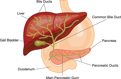
Biliary System
Choledochal cysts are classified into 5 types, based on site of the cyst or dilatation.
- Type I: Most common variety (80-90%) involving saccular or fusiform dilatation of a portion or entire common bile duct (CBD) with normal intrahepatic duct.
- Type II: Isolated diverticulum protruding from the CBD.
- Type III or Choledochocele: Arise from dilatation of duodenal portion of CBD or where pancreatic duct meets.
- Type IVa: Characterized by multiple dilatations of the intrahepatic and extrahepatic biliary tree.
- Type IVb: Multiple dilatations involving only the extrahepatic bile ducts.
- Type V or Caroli's disease: Cystic dilatation of intra hepatic biliary ducts
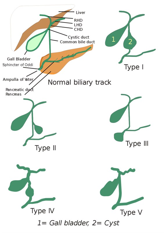
By I, Drriad, CC BY-SA 3.0, https://commons.wikimedia.org/w/index.php?curid=2309266
Epidemiology
Choledochal cysts are rare conditions that are present at birth. It is unclear what causes choledochal cysts.
Surgical Treatment
Bile duct surgery with total cyst removal is the definitive treatment for choledochal cysts. In earlier decades, an operation known as cystenterostomy was performed that only drained the cyst and the bilary reconstruction left the cyst behind. That surgery proved ineffective, leaving many patients with recurrent cholangitis and chronic inflammation in the remaining cyst, and the substantial risk of malignant transformation into cholangiocarcinoma (bile duct cancer).
Today, the surgical treatment for choledochal cysts cyst is complete excision followed by reconstruction of the biliary tree. Future complications include cholangitis and a 2% risk of malignancy, which may develop in any part of the biliary tree. Due to these long-term risks, patients need to be followed regularly to monitor for the development of cholangitis and bile duct cancer.